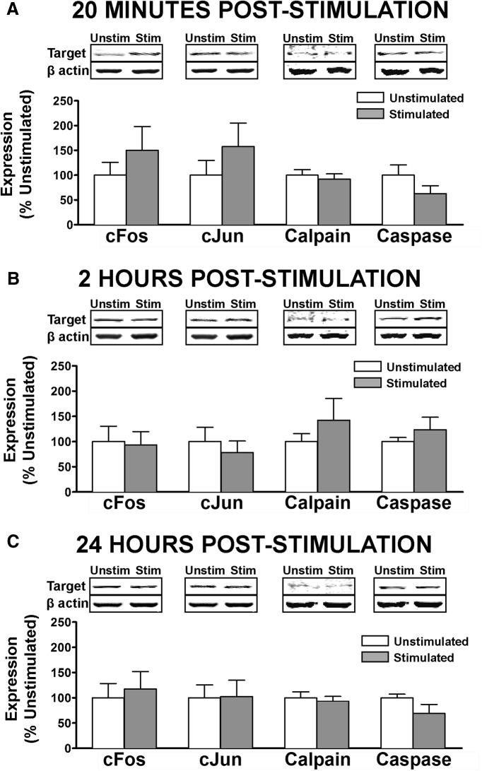
Figure 3.
Assessment of neuronal activity and cell death markers following nociceptive stimulation. Linear Western blot quantification of cell activity and death in cytosolic fractions of ventral lumbar spinal cord, assessed at (A) 20 m, (B) 2 h, or (C) 24 h after intermittent nociceptive stimulation. ANOVA showed no significant increase in broad neuronal activity marker cFos, in other more specific markers of apoptotic cell death (cJun, cleaved caspase3) or in calcium-mediated cell death (calpain I). No significant differences were observed for beta-actin loading control (p > 0.05). Bars represent mean for n = 4 subjects/factorial group (n =12 for INS main effect; n = 8 for time main effect; n = 4 for interaction) with three independent Western blot runs per subject. Error bars represent standard error of the mean.

