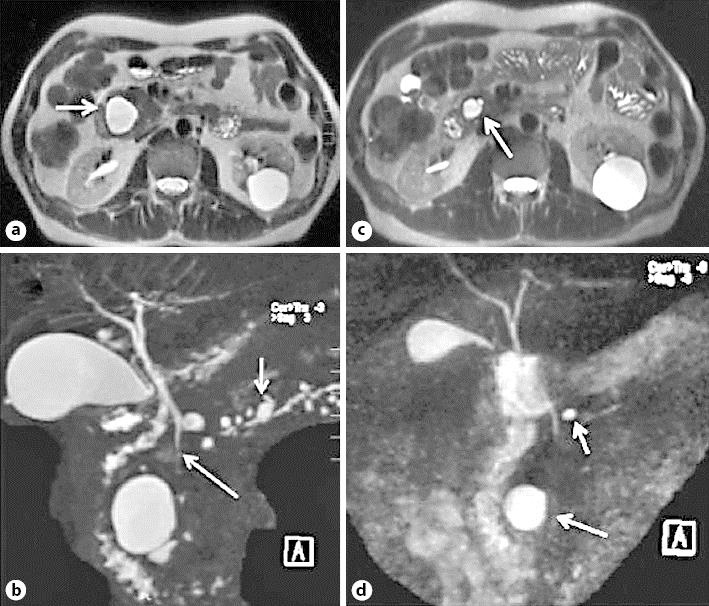
Fig. 2.

MR image 1 year after the initial diagnosis. a, b Signs of atrophy of the pancreatic parenchyma and appearance of multiple cysts, the largest in the head (arrow) and several smaller cystic lesions also in the body and tail (arrows), not communicating with the pancreatic duct. c, d Control images after 6 weeks of corticosteroid treatment showing almost complete regression of the cysts displayed in a and b; only the cyst in the head persists, with a significant decrease in size (arrows).
