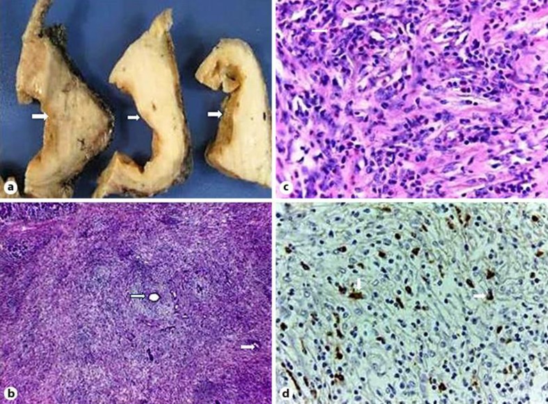
Fig. 4.
a Macroscopic photo showing serial sections of a solid cystic pancreatic lesion, with a thick fibrotic wall (arrows). b Histological picture showing pancreatic atrophy, lymphoplasmacytic infiltration and storiform fibrosis. Some collapsed remnants of small ducts can be seen (arrows). HE. ×40. c Histological picture. Dense inflammatory lymphoplasmacytic infiltration, with some histiocytes and eosinophils (arrows). HE. ×400. d Plasma cells showing positivity for IgG4 (arrows). ×400.

