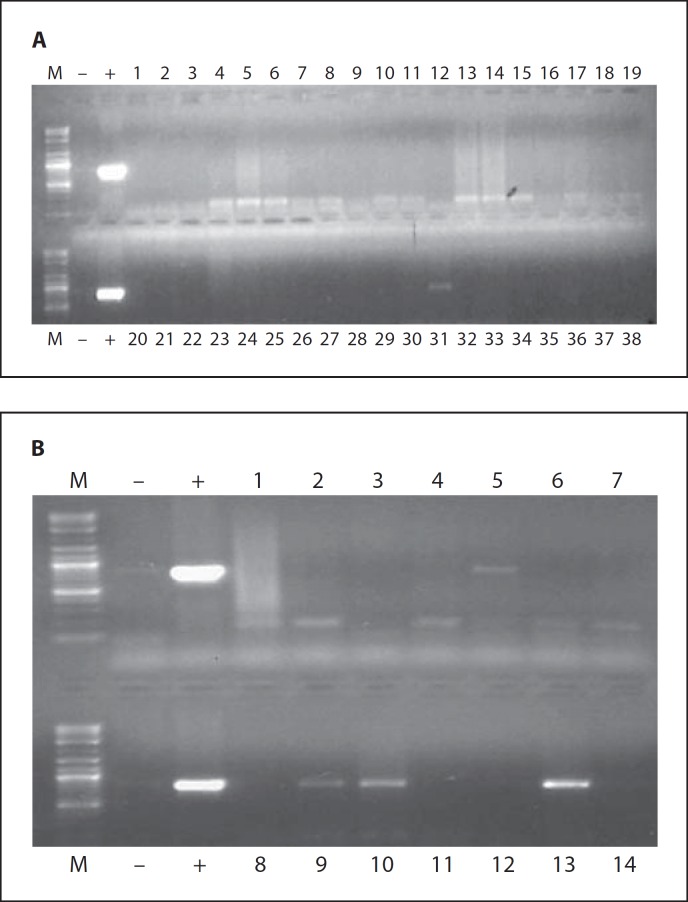
Fig. 7.
Evaluation of Ad-HSV1TK dissemination in Eker rat organs after a single intrauterine leiomyoma injection of 3 β 1010 PFU/cm3 tumor. Total DNA was isolated from tumor tissue and most animal organs as described in the Materials and Methods section. We then performed PCR amplification of the Ad E4 region, and the presence of the E4 region was documented on 1% agarose gel. The 714-bp DNA fragment corresponding to the Ad E4 region was clearly detected in tumor tissues (B10 and B13), while faint bands were detected in 30% of liver (A31 and B5) and 20% of myometrium tissues (B9). The other organs showed negative results. These results are the sum of both Ad-LacZ and Ad-TK groups at 10 days after Ad injection.

