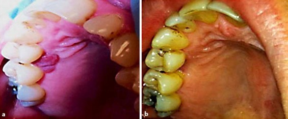
Fig. 2.

Palatal gingiva. a Photograph showing 0.8 × 0.7 cm gingival growth on the palatal aspects between the maxillary bicuspids (before intralesional steroid injection). b Photograph showing disappearance of the lesions 3 weeks after steroid intralesional injection.
