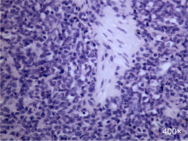
Figure 2.

H&E stain of the biopsy sample.
Notes: There were medium-sized to large cells with finely dispersed chromatin, prominent nucleoli, and abundant eosinophilic cytoplasm (×400).
Abbreviation: H&E, hematoxylin and eosin.

H&E stain of the biopsy sample.
Notes: There were medium-sized to large cells with finely dispersed chromatin, prominent nucleoli, and abundant eosinophilic cytoplasm (×400).
Abbreviation: H&E, hematoxylin and eosin.