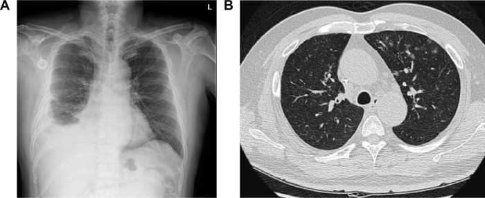
Figure 3.
After steroid administration.
Notes: (A) A chest X-ray shows no obviously increased lung marking, and the implanted port was placed at the right subclavian vein. (B) The repeated chest CT shows marked improvement in ground glass opacity.
Abbreviations: CT, computed tomography; L, left side.

