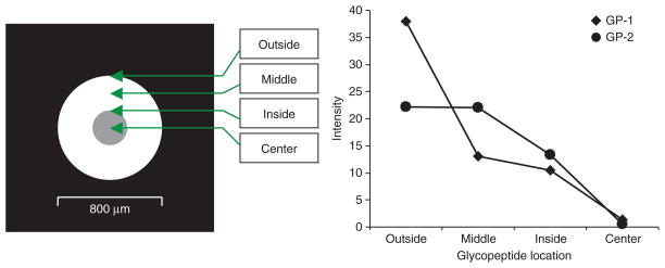
Figure 4.
Distribution of crystallized Gp-1 and Gp-2 glycopeptides in a μFocus plate across the spotted area. The locations where the glycopeptides are distributed in the spot on the plate are indicated by Outside, Middle, Inside, and Center. Gp-1: CGLVPVLAENYN*(5Hex4HexNA c2Sia)K; Gp-2: QQQHLFGSN*(5Hex4HexNAc2Sia) VTDCSGNFCLFR.

