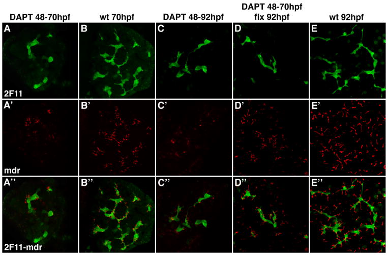
Figure 5. Notch signaling is required for canalicular development.
Confocal projections (5 μm) through the liver of a 70 hpf larva (A) and two 92 hpf (C, D) larvae treated with DAPT from 48 – 70 hpf (A); DAPT from 48 – 92 hpf (C) and DAPT from 48 – 70 hpf (D). A 70 hpf (B) and 92 hpf wild type larva (E) are also shown. All larvae were immunostained with Mdr (red) and 2F11 (green) antibodies. DAPT treatment arrests canalicular development (red) and biliary development (green). Canalicular development reinitiates with DAPT withdrawal (D). Note that the depth of these confocal projections is too small to detect reinitiation of duct development upon DAPT withdrawal.

