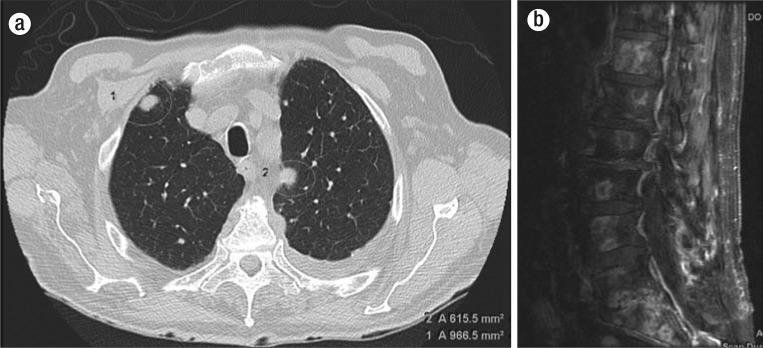
Figure 2.
Imaging. (a) Noncontrast axial CT chest image shows multiple bilateral pulmonary nodules consistent with pulmonary metastatic disease. (b) Postcontrast sagittal T1-weighted MRI of the lumbar spine demonstrates multiple predominantly peripheral enhancing osseous metastases on a background of diffuse marrow signal abnormality.

