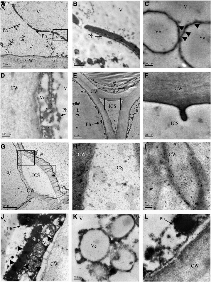
Figure 6.
Subcellular localization of ADE/LAC in litchi pericarp. TEM immunolocalization of the ADE/LAC in the pericarp cells of fruit at day 0 (A–F) and day 4 (G–J) after harvest. A, Ultrastructure of litchi pericarp cells at 0 d exhibited the cytoplasm pressed toward the cell wall by the central large vacuole. In addition, several long Phs were detected in the vacuole. B, A magnified image of the area indicated by the square in A. Numerous gold particles appeared in the long Phs in the vacuole (V). C, A magnified image showing numerous gold particles in the Phs located on the surface of the vesicle (Ve) in the vacuole. D, An additional magnified image showing numerous gold particles in the Phs on the surface of the vesicles close to the cell wall. E, Ultrastructure of litchi pericarp cell at 0 d revealed the cell wall (CW) and intercellular space (ICS). F, Magnified image of the square in E. Few gold particles appeared in the cell wall and intercellular space (ICS). G, Ultrastructure of litchi pericarp cells of 4 d fruit after pericarp browning displayed disintegration of the cell wall. H, A magnified image of the upper square in G. Numerous gold particles in ICS. I, A magnified image of the lower square in G. Numerous gold particles were detected in the disintegrated cell wall. J, Fewer gold particles were detected in Phs after pericarp browning at 4 d than in those at 0 d. K and L, Control images without anti-ADE/LAC incubation showed no gold particles.

