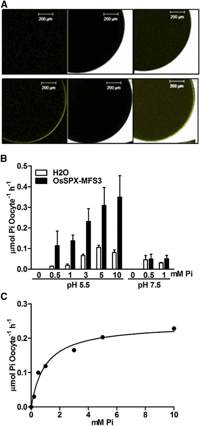
Figure 4.
A, Subcellular localization of OsSPX-MFS3 in X. laevis oocytes. cRNAs of nYFP-fused OsSPX-MFS3 were injected into oocytes and incubated for 48 h in BS before imaging with confocal laser scanning microscopy. B, Pi influx activity of OsSPX-MFS3. Oocytes were incubated for 48 h in BS (without Pi) and then transferred into BS solution containing different Pi concentrations containing 32P (2 mCi mL–1) for 1 h. The radioactivity response to increasing concentrations of externally applied Pi was recorded at pH 5.5 and 7.5. Mean ± se of the mean (n = 10 oocytes) is shown. C, Nonlinear regression of Pi uptake of OsSPX-MFS3 versus external Pi concentration at pH 5.5 was used to estimates the Km value. Oocytes were incubated for 48 h in BS (without Pi) and then transferred into BS solution containing different Pi concentrations containing 33P (5 mCi mL–1) for 1 h. The radioactivity response to increasing concentrations of externally applied Pi was recorded at pH 5.5. Estimated Km and Vmax were 1.101 ± 0.03 mm and 0.24 ± 0.01 μmol Pi oocyte–1 h–1, respectively. Bars = 200 µm.

