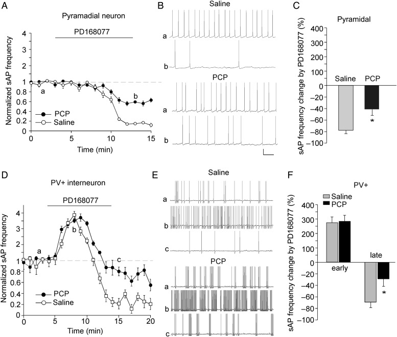
Figure 8.
The effects of D4R on sAP frequency in PFC pyramidal neurons and PV+ interneurons were altered in the PCP model of schizophrenia. (A,D) The plot of normalized sAP frequency showing the effect of PD168077 in pyramidal neurons (A) and PV+ interneurons (D) from saline- versus PCP (5 mg/kg, 3-day)-injected mice. (B,E) Representative sAP traces at different time points (denoted by a–c in plots A,D) in pyramidal neurons (B) and PV+ interneuron (E) from saline- versus PCP-injected mice. Scale bars: 20 mV, 1 s. (C,F) Bar graph summary of the percentage change in sAP frequency by PD168077 in pyramidal neurons (C) and PV+ interneurons (F) from saline- versus PCP-injected mice. *P < 0.05.

