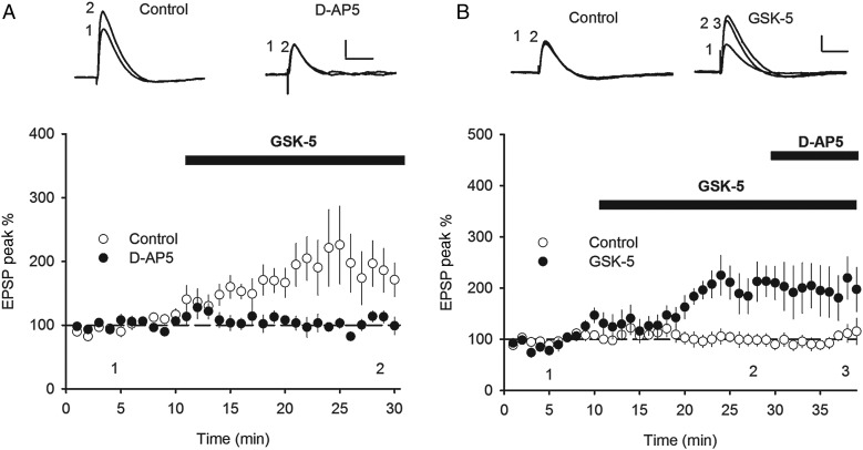
Figure 5.
M1R-induced EPSP enhancement is NMDAR dependent. (A) The NMDAR antagonist D-AP5 (50 µm) prevented the increase in EPSP amplitude induced by application of GSK-5 (500 nm) in whole-cell current clamp recordings. (B) Application of D-AP5 (50 µm) after GSK-5 (500 nm) failed to reverse the increase in EPSP amplitude induced by GSK-5. Data plotted as mean ± s.e.m. Example voltage traces in response to synaptic stimulation taken from Points 1 or 2 as indicated. Scale bars: 2 mV and 50 ms (A,B).

