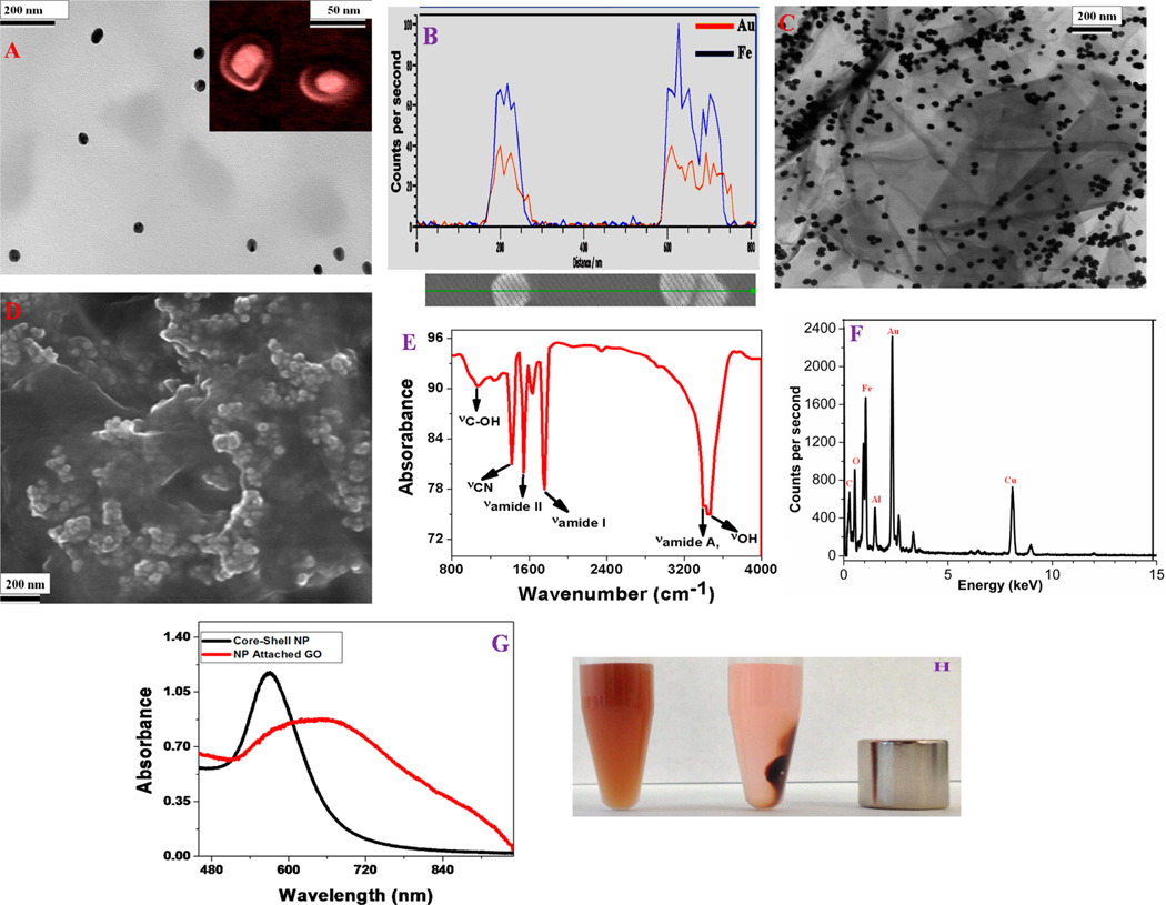
Figure 1.
(A) High-resolution TEM image using JEM-2100F transmission electron microscope showing the morphology of iron magnetic core–gold plasmonic shell nanoparticles; (inset) high-resolution SEM picture confirming the core–shell morphology of freshly prepared nanoparticles. (B) EDX mapping data of freshly prepared core–shell nanoparticle showing the presence of Fe and Au. (C) High-resolution TEM picture showing the morphology of freshly prepared core–shell nanoparticle attached multifunctional hybrid graphene oxide. (D) High-resolution SEM picture showing the three-dimensional view of hybrid graphene oxide, which clearly shows the formation of core–shell nanoparticle assembly on graphene oxide surface. (E) FTIR spectrum from freshly prepared core–shell nanoparticle attached multifunctional hybrid graphene oxide showing the existence of amide A, I and II bands, as well as –CN band, which indicate the formation of amide bond. The stretches –OH and –C–OH groups due to the graphene oxide also be seen on the FTIR spectra. (F) EDX data of freshly prepared multifunctional hybrid graphene oxide showing the presence of Fe, Au, C, and O. We have also observed Cu and Al peaks in the EDX data, which originate from the support grid. (G) Extinction spectra of core–shell nanoparticle and nanoparticle attached hybrid graphene oxide. Due to the formation of core–shell nanoparticle assembly on graphene oxide surface, the excitation spectra is very broad for hybrid material. (H) Photograph showing that the core–shell nanoparticle attached hybrid graphene oxide is highly magnetic, which allows them to be separated by using a bar magnet.

