
FIGURE 1:
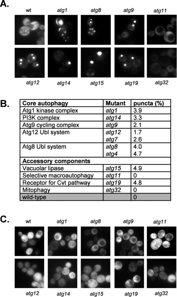
[PSI+] prions are formed in autophagy mutants. (A) Representative fluorescence micrographs for the wild-type and indicated autophagy mutant strains containing the Sup35NM-GFP plasmid. The Sup35NM-GFP plasmid was induced for 1 h using copper before visualizing aggregate formation after 16 h of growth. (B) The aggregation frequency was calculated in the indicated autophagy mutants as a percentage of the number of cells containing fluorescent foci out of ∼300 cells counted. (C) Representative fluorescence micrographs for strains cured with GdnHCl.