
FIGURE 2:
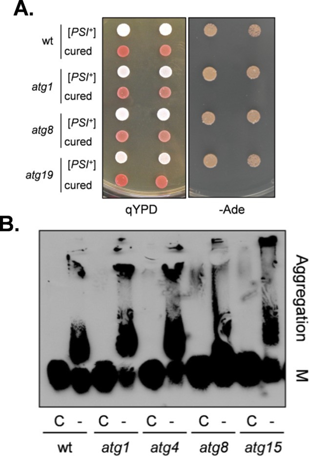
The [PSI+] prion is formed in autophagy mutants. (A) [PSI+] prion formation was visualized in the wild-type (74D-694) and atg1, atg8, and atg19 mutant strains by pink/white colony formation and growth on minimal medium in the absence of adenine. Curing with GdnHCl gives rise to red Ade− colonies, confirming the de novo formation of [PSI+] in these cells. (B) Cell extracts were prepared from exponentially growing cells and analyzed by SDD-AGE. SDS-resistant Sup35 aggregates were detected in atg1, atg4, atg8, and atg15 mutant strains. Aggregate and monomer (M) forms are indicated.