
FIGURE 6:
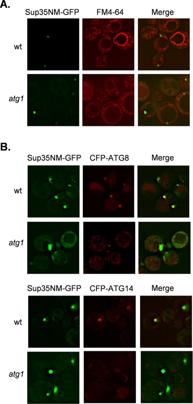
Sup35 aggregate formation occurs at similar intracellular sites in wild-type and atg1 mutant strains. (A) The vacuolar dye FM4-64 was used to visualize vacuolar membranes in wild-type and atg1 mutant strains. Similar vacuolar Sup35-GFP foci were detected in the wild-type and atg1 mutant strains after copper induction of the SUP35NM-GFP fusion construct for 1 h. (B) CFP-ATG8 and CFP-ATG14 were used as markers of PAS in wild-type and atg1 mutant strains. Strains were grown for 48 h in the presence of 4 mM spermidine to induce autophagy and SUP35NM-GFP induced with copper for 1 h. Sup35 aggregates form adjacent to PAS markers in the wild-type strain.