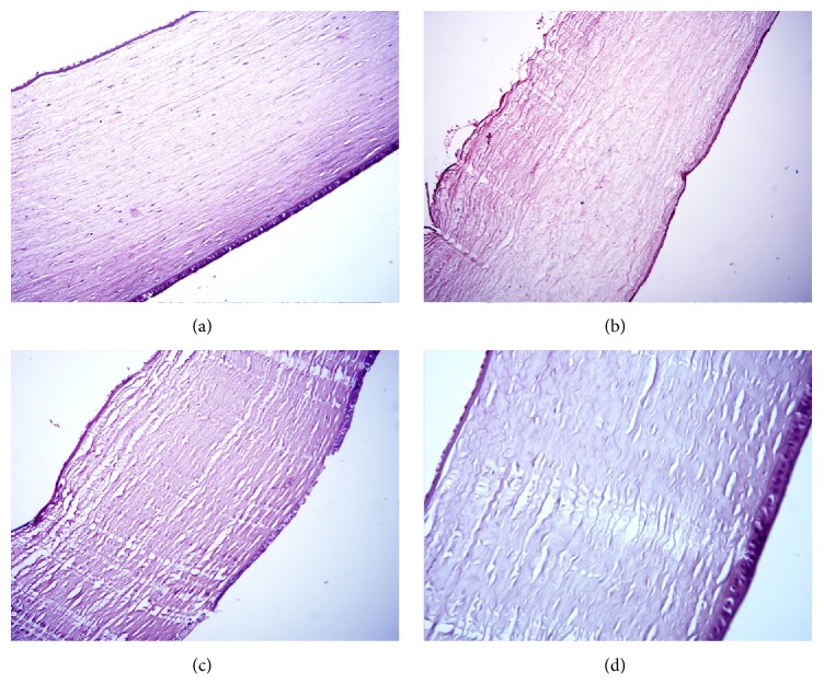
Figure 7.
Histology picture of the cornea, stained with hematoxylin and eosin, magnification ×200, on day 4 of uveitis. (a) Control (n = 2): cornea is lined with epithelium and endothelium; collagen fibers and keratocytes (specialized corneal fibroblasts) are visible; (b) placebo (n = 6): swelling and loosening of corneal collagen fibers, endothelial desquamation, partial destruction of Descemet's membrane; epithelium is not changed; (c) SOD1 (n = 6): moderate loosening of the corneal collagen fibers, partial endothelial desquamation, cell infiltration is absent; (d) SOD1 nanozyme (n = 6): normal structure of the cornea.

