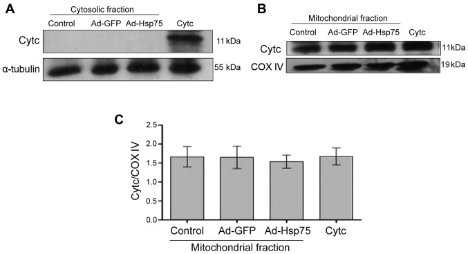
Figure 7.
Lack of cytochrome c (Cytc) protein expression in the cytosolic fraction. (A) The Cytc in the cytosolic fraction was analyzed by western blot analysis. α-tubulin was used as an internal control for cytosolic protein. (B) Cytc in the mitochondria was analyzed by western blot analysis. COX IV protein served as an internal control for mitochondrial protein. (C) Quantitative analysis of Cytc protein expression in mitochondria. Data represent the means ± SEM. Three independent experiments were performed. Ad-GFP, adenovirus green fluorescent protein; Ad-Hsp75, adenovirus heat shock protein 75.

