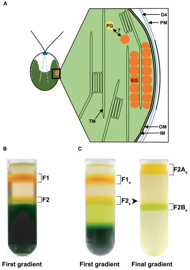
FIGURE 1.
Schematic drawing of the eyespot in Chlamydomonas reinhardtii and distribution of different fractions enriched in eyespots after flotation on discontinuous sucrose gradients. (A) Schematic longitudinal section through the eyespot in the region of the four stranded microtubular root (D4), which is important for eyespot positioning. The functional eyespot consists of local specializations of different compartments. The part of the plasma membrane (PM) overlying the eyespot globules (EG) is thickened and in close association with the inner and outer chloroplast envelope (IM/OM). The EG are in contact with the thylakoid membrane (TM) and arranged in layers, the outermost being also in contact with the chloroplast envelope. The possible link between plastoglobules (PG) and EG is indicated. (B) Separation of the cell homogenate in the first gradient using the established eyespot isolation method by Schmidt et al. (2006). (C) Separation of the cell homogenate in the first gradient using the here described (see Materials and Methods) modified cell rupture method for an extended (“e”) eyespot fraction; separation of fraction 2 (F2e) from these gradients in fraction 2A (F2Ae) and 2B (F2Be) in the final gradient. Note the increased amount of F2e and the decreased amount of F1e and thylakoid debris in comparison to the original cell rupture method.

