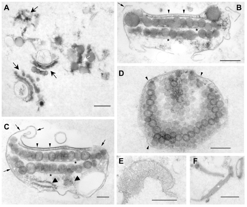
FIGURE 2.

Characterization of the extended eyespot fraction 2A (F2Ae) by transmission electron microscopy revealed enrichment of intact eyespots. Overview (A) and details (B–F). (A) Black arrows indicate well-preserved eyespots, whereas the white arrow indicates structurally less well-preserved eyespots with fused EG; scale bar: 600 nm. (B,C) Cross-sections through isolated, double-layered eyespots associated with eyespot and other membranes. The globule layers are subtended by a thylakoid (black asterisk). Small arrowheads: plasma membrane area overlying the EG. Note the close association with the chloroplast envelope membranes and the thickened, electron-dense appearance of the plasma membrane in this region. In addition, the fuzzy fibrillar material typically observed in situ between the plasma membrane and chloroplast envelope in the region of the eyespot apparatus (Kreimer, 2009) is preserved. Small arrows point to normal plasma membrane regions tending to enwrap the globule layers and not eyespot-associated structures like a thylakoid stack (white asterisk) or ribosomes (large arrowheads); scale bars: 150 nm. (D) Tangential section through a globule layer. Arrowheads indicate membranes enclosing the eyespot; scale bar: 250 nm. (E,F) Co-isolated free mitochondrial fragment (E) and free thylakoid stack (F); scale bars: 400 nm.
