
Figure 1.
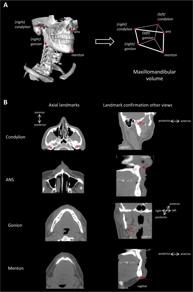
Analysis of maxillomandibular volume from computed tomography scans of the head. A maxillomandibular volume was calculated from the polyhedral volume made of vectors connecting points of the left and right condyle, left and right gonion, anterior nasal spine (ans), menton. (A) Three-dimensional reconstruction of the craniofacial skeleton showing visible craniofacial landmarks. The three dimensional coordinates were used to construct a polyhedral volume as shown. (B) Craniofacial landmarks were marked on axial image slices using the brush tool of the imaging software and centroid coordinates (x,y,z) of each landmark exported into a customized spreadsheet for volume calculation. Axial slices are shown for each landmark with corresponding sagittal or coronal views to confirm correct placement of each landmark.