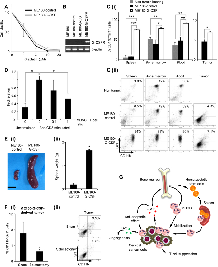
Figure 2. MDSC accumulation in G-CSF-producing cervical cancer derived tumor bearing mice.
(A) In vitro sensitivity of cervical cancer cells to cisplatin. ME180-G-CSF or ME180-control cells were exposed to the indicated dose of cisplatin for 72 hours. Cell viability was assessed using the MTS assay. Data points, mean values; bars, SD. Data are shown as the means of triplicate samples. (B) RT-PCR analysis of the G-CSFR and β-actin mRNA levels of the cervical cancer cells. ME180-G-CSFR cells, into which a G-CSFR expression vector had been stably transfected, were used as positive controls. (C) CD11b+ Gr-1+ cell populations in G-CSF-producing cervical cancer. Mice were inoculated with ME180-G-CSF (n = 6), ME180-control cells (n = 6), or PBS alone (n = 6). Four weeks after the inoculation, their spleen, bone marrow, blood, and tumors were collected. (i) CD11b+ Gr-1+ cell populations were counted by flow cytometry. Bars, SD. *P < 0.05, **P < 0.01, ***P < 0.001, Two-sided Student’s t test. (ii) Representative dot plot. The percentage of the CD11b+ Gr-1+ cell is indicated. (D) Ability of G-CSF-induced CD11b+ Gr-1+ cells to suppress anti-CD3 mAb-stimulated T cells. Balb/c mice were subcutaneously treated with 10 μg recombinant human G-CSF for three days. CD11b+ Gr-1+ cells were isolated from their spleen. CD8+ T cells (2 × 10⁵ cells/well) were isolated from syngeneic mice and co-cultured with CD11b+ Gr-1+ cells at indicated ratios. Cells were incubated for 72 hours, after which BrdU was added for an additional 24 hours. T cell proliferation was determined by BrdU incorporation. Bars, SD. *P < 0.05, Two-sided Student’s t test. (E) Splenomegaly in G-CSF-producing cervical cancer. Mice were inoculated with ME180-G-CSF (n = 6) or ME180-control cells (n = 6). Their spleens were collected 4 weeks later. (i) Representative photos of the spleen (bar = 1 cm). (ii) Spleen weight. Bars, SD. *P < 0.05, Wilcoxon rank sum test. (F) The effect of splenectomy on MDSC accumulation in ME180-G-CSF-derived tumors. Mice that underwent splenectomy (n = 5) or sham surgery (n = 5) were inoculated with ME180-G-CSF. Three weeks after the inoculation, their subcutaneous tumors were collected. (i) CD11b+ Gr-1+ cell populations in tumors. Bars, SD. *P < 0.05, Two-sided Student t test. (ii) Representative data. (G) Proposed paracrine mechanism responsible for the chemoresistance in cervical cancer.

