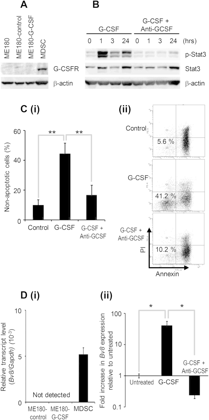
Figure 3. Mechanism of MDSC-mediated cisplatin resistance.
(A) Western blot analysis of G-CSFR and β-actin expression in MDSC. (B) The effect of G-CSF on Stat3 activation of MDSC. MDSC were treated with 10 ng/mL G-CSF in the presence or absence of 100 μg/mL anti-G-CSF-neutralizing antibody. (i) Cells were cultured for indicated time and then activation of Stat3 in MDSC was assessed by Western blotting. (C) The effect of G-CSF on the survival of MDSC. MDSC were treated with 10 ng/mL G-CSF in the presence or absence of 100 μg/mL anti-G-CSF-neutralizing antibody. (i) Cells were cultured for 24 hours and then assayed for apoptosis by flow cytometry. The pooled data indicating the non-apoptotic cells were shown. Bars, SD. **P < 0.01, Two-sided Student’s t test. (ii) Representative dot plot. The percentage of the non-apoptotic cells is indicated. (D) The effect of G-CSF on the expression of Bv8 in MDSC. (i) Cervical cancer cells and MDSC were harvested, and their Bv8 expression was assessed by real-time RT-PCR. The expression level of Bv8 mRNA was normalized to that of GAPDH mRNA. (ii) MDSC were treated with 10 ng/mL G-CSF in the presence or absence of 100 μg/mL anti-G-CSF-neutralizing antibody for 4 hours. Then their Bv8 expression was assessed. Bars, 95% confidence interval. *P < 0.05, Two-sided Student’s t test.

