


Figure 2.
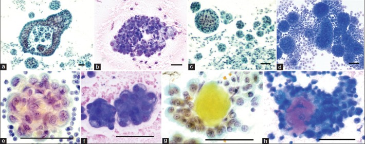
Cytomorphology of epithelioid mesothelioma cells and tissue fragments in effusions. The tissue fragments sometimes show gaps or windows, (a and b) occur both as spheres with smooth surfaces (c and d) and in berry-like clusters with scalloped surface (e and f). The tissue fragments may contain acidophilic extracellular matrix cores also known as collagen or basement membrane cores (g and h) which with May-Grünewald-Giemsa (MGG) becomes strongly acidophilic, similar to the extracellular granular material that can be seen in the background [Figures 1 and 2f], indicating large amounts of hyaluronan (left PAP stain, right MGG stain; bar = 50 μm). Photo: VA frames a, c, e and g; AH frames b, d, f and h