
Fig. 2.
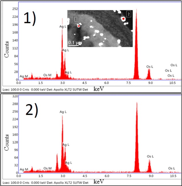
EDS spectra of silver signal 1 inside the yeast cell and 2 on the outer CW. TEM micrograph shows distribution of AgNPs inside the planktonic cell and also on the surface of the outer CW after 24 h incubation. EDS spectra of silver nanoparticles demonstrate the presence of elemental silver signal in the sample