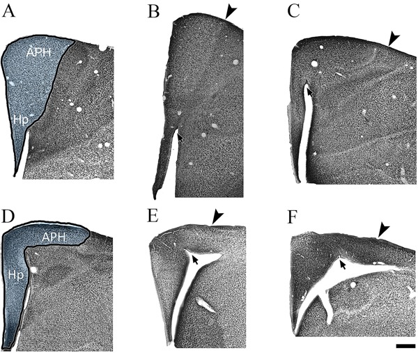
Figure 2. Hippocampal neurons of Actitis macularia. Coronal series of NeuN-immunolabeled sections of the A. maculariahippocampal formation. The left to right sequence is from the frontal to the occipital pole of the hippocampal formation. In the first sections of the top and bottom rows, the dark line defines the area of interest. The arrowheads indicate limits of the area of interest. The arrows indicate the paraventricular sulcus. APH: parahippocampal area; Hp: hippocampus. Scale bar: 500 µm.

