
Fig. 5.
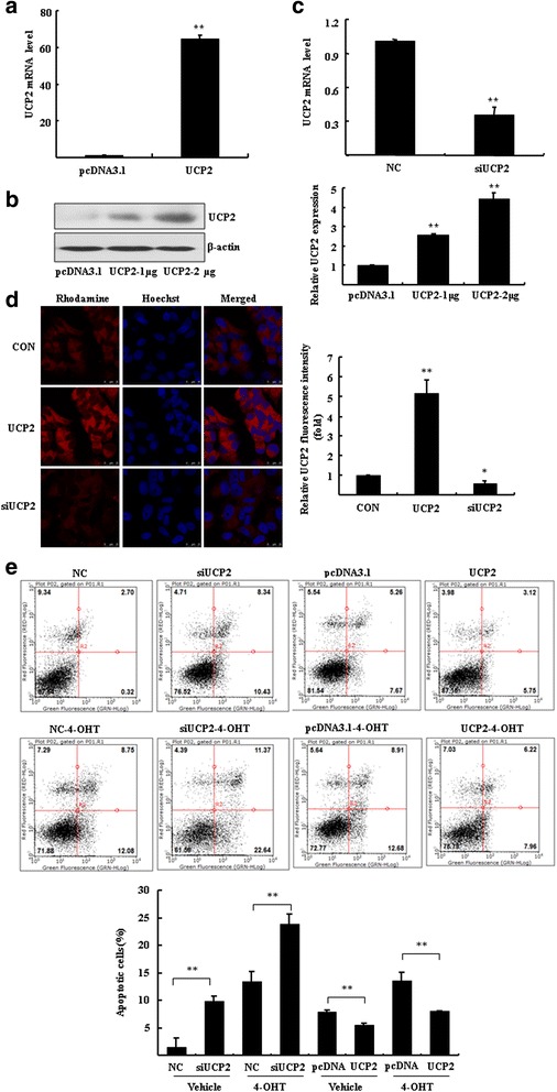
UCP2 modulated the 4-OHT/FUL-induced apoptosis in breast cancer cells. a-c MCF7 cells were transfected with UCP2 plasmid and empty vector pcDNA3.1 or 200 nM siRNA of UCP2 for 48 h. RT-qPCR and Western blotting were performed to determine the expression of UCP2. The ratio UCP2/β-actin is quantified (**P < 0.01 vs. empty vector control). d The immunofluorescence assay was performed to analyze the expression and location of UCP2 in MCF7 cells. e Annexin V-PI staining assay was performed to determine apoptosis in MCF7 cells. Bar graphs indicated the percentage of apoptotic cells. **P < 0.01 vs. negative control (NC) or empty vector control (pcDNA3.1)