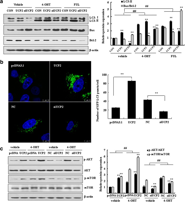
Fig. 6.

UCP2 modulated autophagy through activation of the PI3K-Akt-mTOR pathway. a MCF7/LCC9 cells were transfected with UCP2 plasmid or siRNA of UCP2 for 48 h. Western blotting was performed to analyze the expression of cleaved LC3 and the ratio of Bax/Bcl-2. Bar graphs indicated the relative levels of LC3-II and ratio of Bax/Bcl-2 normalized to β-actin. *P < 0.05, **P < 0.01 vs. control. ## P < 0.01 vs. vehicle control. b MCF7 cells were cotransfected with GFP-LC3 together with UCP2 plasmid or 200 nM siRNA of UCP2 for 48 h. Cells were imaged under a confocal microscopy (scale bar = 10 μm). Numbers of GFP-LC3 puncta per cell were counted (**P < 0.01 vs. empty vector control or negative control, n = 10). c Western blotting was performed to analyze p-Akt, Akt, p-mTOR, mTOR in MCF7/LCC9 cells. β-actin was served as loading control. Bar graphs indicated the relative levels of p-Akt/Akt and p-mTOR/mTOR normalized to β-actin. ** P < 0.01 vs. empty vector control or negative control, ## P < 0.01 vs. vehicle control
