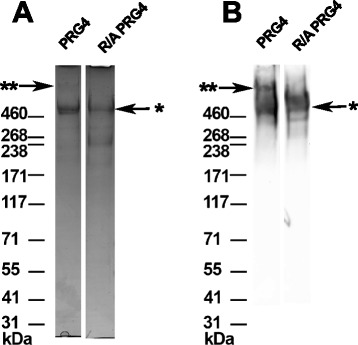
Fig. 1.

Characterization of PRG4 and reduced and alkylated (R/A) PRG4. Protein stain on 3-8 % Tris-Acetate SDS-PAGE stained with SimplyBlue SafeStain (a) and immunoreactivity with anti-PRG4 antibody 5C11 (b). ** and * indicate high MW multimeric species and monomeric PRG4 species, respectively, with the former being present in the non-reduced samples and absent from the R/A PRG4
