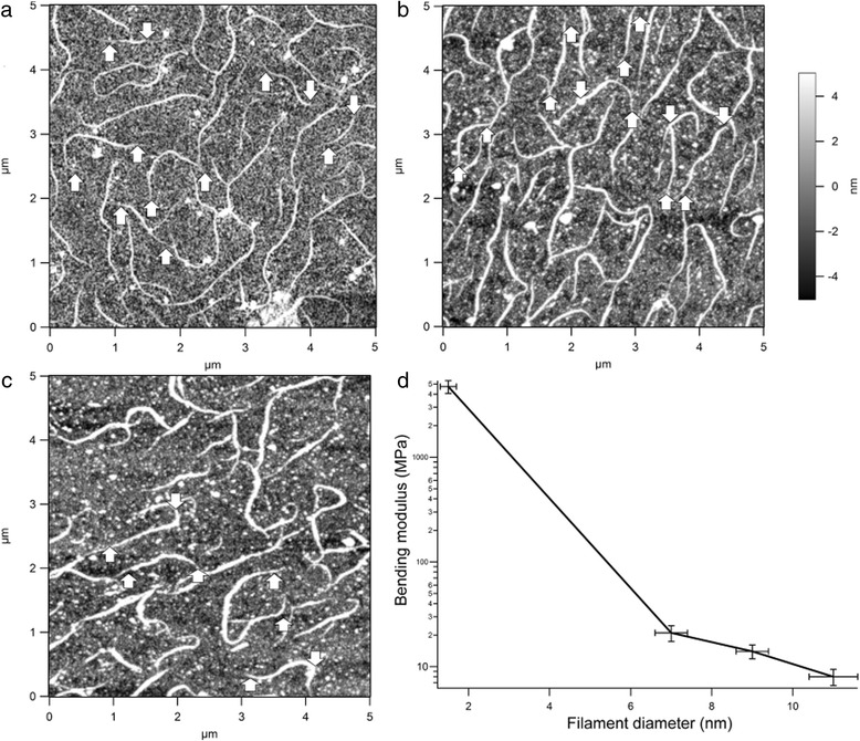
Fig. 8.

Atomic force microscopy analysis of type II collagen fibrillogenesis. a-c Images of collagen fibrils formed after a 10 min, b 20 min, and c 30 min of incubation. The upward pointing arrows show tapered ends and downward pointing arrows show blunt ends. d Bending modulus versus filament diameter extracted from AFM images at different time points of the fibrillogenesis process
