
Figure 1.
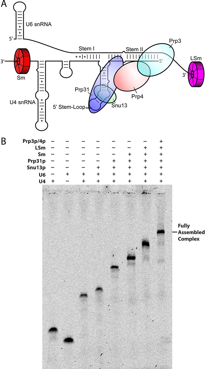
(A) Secondary structure representation of the yeast U4/U6 di-snRNP. Each snRNP protein is color-coded and labeled accordingly. (B) Stepwise assembly of the full U4/U6 di-snRNP under sub-stoichiometric conditions followed by electrophoretic mobility shift assay (EMSA). Consecutive binding of each protein results in complete gel shifts, indicating step-wise assembly of the snRNP.