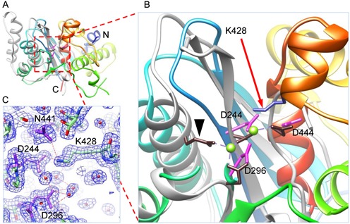
Figure 1.

Structure of wild-type gp2C. (A) and (B) The gp2C structure (ribbon diagram rainbow-colored from the N-terminus to the C-terminus) is superimposed with that of B. halodurans RNase H (grey; RCSB PDB code 2G8H). Side chains of acidic residues in the active site of gp2C and K428 are shown as stick models in magenta and blue, respectively. The active site residues of RNase H are shown as stick models in brown. One of the active site residue of RNase H, E109, is indicated with a black arrow head. The two bound Mg2+ in RNase H are shown as green spheres. (C) The 1.55 Å resolution 2mFo–DFc electron density map of gp2C contoured at 1.0 sigma superimposed with the refined model. Water molecules, spheres in magenta. H-bonds, dashed lines in red.
