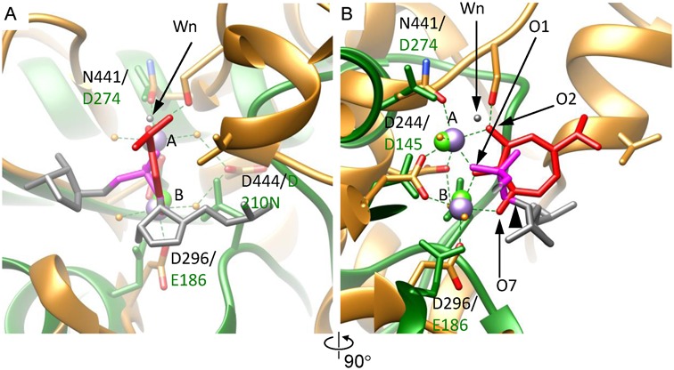
Figure 5.

The metal ion binding mode of gp2C–K428A:Mn2+:BTP mimics the DNA-binding state. (A) The gp2C–K428A:Mn2+:BTP structure (gold) is superimposed onto human RNase H1 in complex with an RNA/DNA hybrid and Ca2+ (RCSB PDB code 2G8H) (green) assuming the metal ion binding modes are conserved and the bound BTP reflects the DNA scissile phosphate (see text). Side chains of active side residues are shown as stick models. Each superimposed pair of active site residues are indicated with the residue numbers in gp2C on the top and in RNase H1 on the bottom, respectively. The two Mn2+ in gp2C–K428A structure are shown as purple spheres and labeled with A and B, respectively. The two Ca2+ ions in RNase H1 are shown as green spheres. The RNA strand of the RNA/DNA hybrid in the RNase H1 structure is shown as stick models in grey. For clarity, only two nucleotides are shown. The scissile phosphate is in magenta. The leaving 3′-OH group is indicated with a black arrowhead in (B). The nucleophilic water in the RNase H1 structure is indicated with a black arrow and labeled Wn. The oxygen atoms O1, O2 and O7 of BTP are indicated. (B) 90° about the vertical axis from (A).
