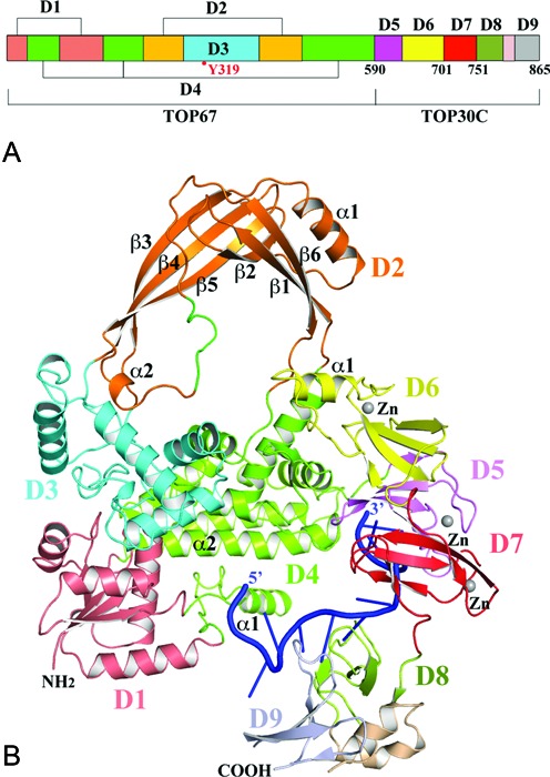
Figure 1.

Structure of full-length E. coli topoisomerase I (EcTOP1) in complex with single-stranded DNA (ssDNA). (A) Domain arrangement of E. coli topoisomerase I. Between D8 and D9, there is a helical hairpin. (B) A ribbon diagram of full-length EcTOP1 in complex with ssDNA. Full-length EcTOP1 includes four N-terminal domains: D1 (deep salmon), D2 (orange), D3 (cyan) and D4 (green); and five C-terminal domains: D5 (pink), D6 (yellow), D7 (red), D8 (lime) and D9 (grey). The helical hairpin between D8 and D9 is colored in wheat. A ssDNA that binds to the C-terminal domains is colored in blue. Each Zn(II) is represented as a gray sphere. The secondary structures of D2 and part of D4 and D6 are labeled for discussion purposes. A part of the loop (colored in green) between α2 and β6 of D2 includes a charged and conserved sequence of R442KGDEDR, which is highly flexible and was not observed in earlier TOP67 structures. Figures 1B, 2, 4 and 7A are prepared with the program PyMOL (http://www.PyMOL.org).
