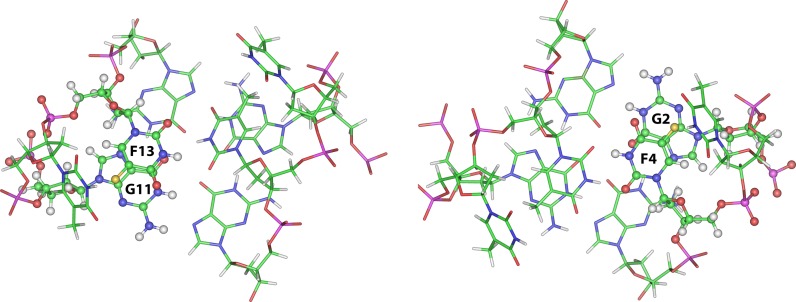
Figure 4.
Bottom view representations of the molecular models of the quadruplexes formed by TBA-F13 (left) and TBA-F4 (right). The structures are oriented with the 5′ and 3′-ends upward. The TGT loop and the G1-G6-G10-G15 tetrad have been omitted for clarity. Backbones and bases are depicted in coloured ‘stick’ (carbons, green; nitrogens, blue; oxygens, red; hydrogens, white; fluorine, yellow). The F13 (left) and F4 (right) residues and the respectively overlying G11 and G2 are reported in ball and sticks, in order to point out the effectiveness of the stacking interaction between them.

