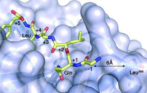
Figure 1.

Crystal structure of the β-clamp bound to a polymerase II peptide. Surface representation of the E. coli β-clamp bound to the β-binding motif of polymerase II (color-coded sticks, PDB ID 3D1E). β has a well-defined groove near its C-terminus where partners interact. The conserved Gln and Leu residues at positions +1 and +4 of the β-binding motif are labeled. The C-terminal residue (Leu366) of β is also labeled and the distance to the N-terminal amino acid of the peptide is indicated.
