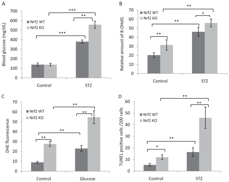
Fig. 1.
Diabetes induced marked oxidative stress and apoptosis in Nrf2 KO cardiomyocytes. A, B, and D, WT and Nrf2 KO mice (8 weeks old, male) were given STZ (150 mg/kg b.wt.) or 0.1 M citrate buffer (vehicle control) intraperitoneally. Mice were sacrificed, and heart tissues were examined 2 weeks after STZ injection. A, blood glucose level. Tail blood glucose levels were measured at the end of the experiment (n = 8). B, 8-OHdG formation. Oxidative DNA damage was assayed by immunofluorescent staining of 8-OHdG in heart sections. C, ROS production. AMVM were isolated from WT and KO mouse hearts, treated with glucose (30 mM, 18 h), and stained with DHE for production of ROS. Fluorescence intensities were quantified and are shown as means and S.D. from four to six samples. D, apoptosis. Apoptosis was measured by using the DeadEnd Fluorometric TUNEL assay of heart sections. *, p < 0.05; **, p < 0.01; ***, p < 0.001.

