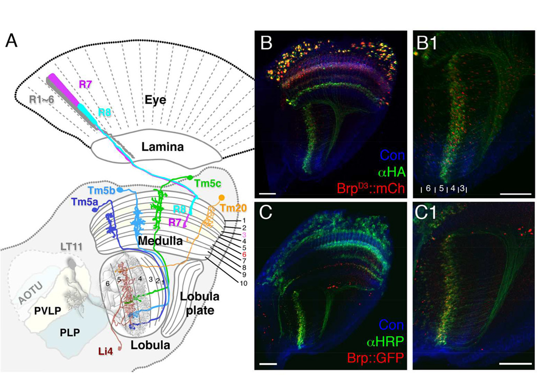
Figure 1.
The axons of chromatic Tm neurons, Tm5a/b/c and Tm20, form terminals in deep lobula strata. (A) Schematic illustration of the Drosophila visual system, including the compound eye’s retina, and successive optic neuropils, the lamina, medulla and lobula, with central brain regions. R1–6 innervate the lamina, and R7, R8 the medulla. A single lobula projection neuron LT11 (gray) receives chromatic inputs from strata Lo4 and Lo5 strata and projects an axon to optic glomeruli in the posterior ventrolateral protocerebrum (PVLP). A representative lobula intrinsic neuron (Li4, brown) elaborates web-like processes in lobula stratum Lo5. Its cell body is located primarily in the posterior lateral cell body region (LCBR). AOTU, anterior optic tubercle; PLP, posterior lateral protocerebrum. (B, B1) ortC1a-LexA::VP16 drives expression of a HA-tagged membrane marker (αHA, green) and an active zone marker, Brp::mCherry (red) in the chromatic Tm neurons. Presynaptic sites of Tm5a/b/c and Tm20 are located in the deep lobula strata (Lo4, Lo5, and sparsely in Lo6). (C,C1) The presynaptic sites of chromatic Tm neurons marked using the STaR method, which expresses GFP-tagged Brp (pseudo-colored red) from the endogenous brp promoter (see main text for details), showing a pattern of presynaptic sites similar to (B1) in the lobula. B1 and C1 enlarged from B and C, respectively. Scale bar, 20 µm for B-C1.

