Figure 2.
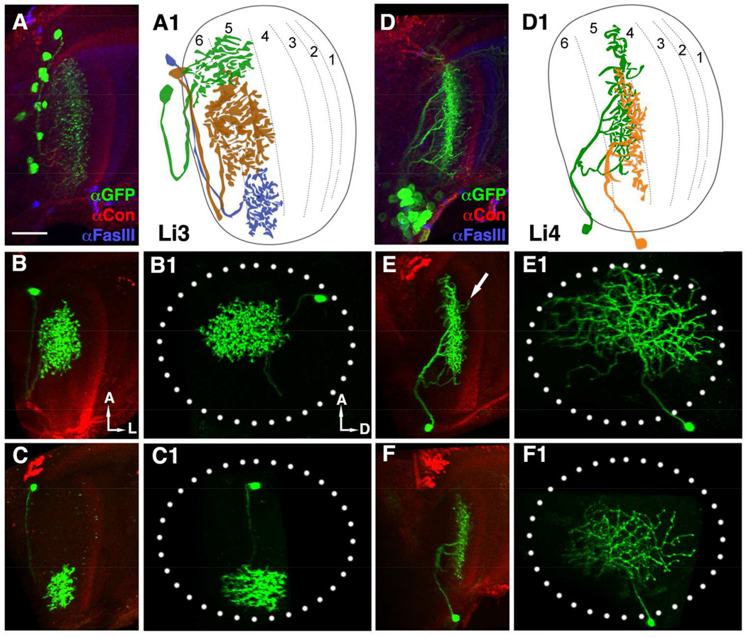
Lobula intrinsic neurons, Li3 and Li4, extend dendritic arbors in deep strata of the lobula. (A, D) Li3- and Li4-specific Gal4 drivers drive expression of a membrane-tethered GFP marker (mCD8::GFP, green) to reveal the dendritic arbors of these cells. Anti-Fas III (blue) and anti-Connectin (magenta) immunolabeling were used to mark lobula strata 1, 2, 4, 5 and 3, respectively (Gao et al., 2008). In each optic lobe, about 12 Li3 neurons elaborate dendritic arbors spanning the entire lobula strata 5 and 6 (A). Approximately 30 Li4 neurons, clustered in the posterior lobula cortex have dendritic arbors covering the entire lobula stratum 5.
(A1 and D1) Schematic drawings of Li3 (A1) and Li4 (D1) neurons.
(B-C1, E-F1) Single Li3 (B-C1) or Li4 (E-F1) neurons labeled with mCD8::GFP using the single-cell flip-out technique and visualized in side (B,C,E,F) and plan views (B1,C1,E1,F1) by anti-GFP (green). (B-C1) Each Li3 neuron elaborates a dense dendritic arbor spanning an area corresponding to about 11–20% of the visual field (dotted ellipse, B1 and C1) in lobula strata 5 and 6 (B and C). (E-F1) Each Li4 neuron extends a dendritic arbor that covers a large portion (45–78%) of the visual field (dotted ellipse) in lobula stratum 5 (E1, F1). Occasional distal branches of Li4 invade the neighboring lobula stratum 4 (arrow, E).
(A, B, C, D, E, F) ventral views; (B1, C1, E1, F1) frontal views. A: anterior; L: lateral; D: dorsal. Scale bar: 20 µm in A for A-F1.

