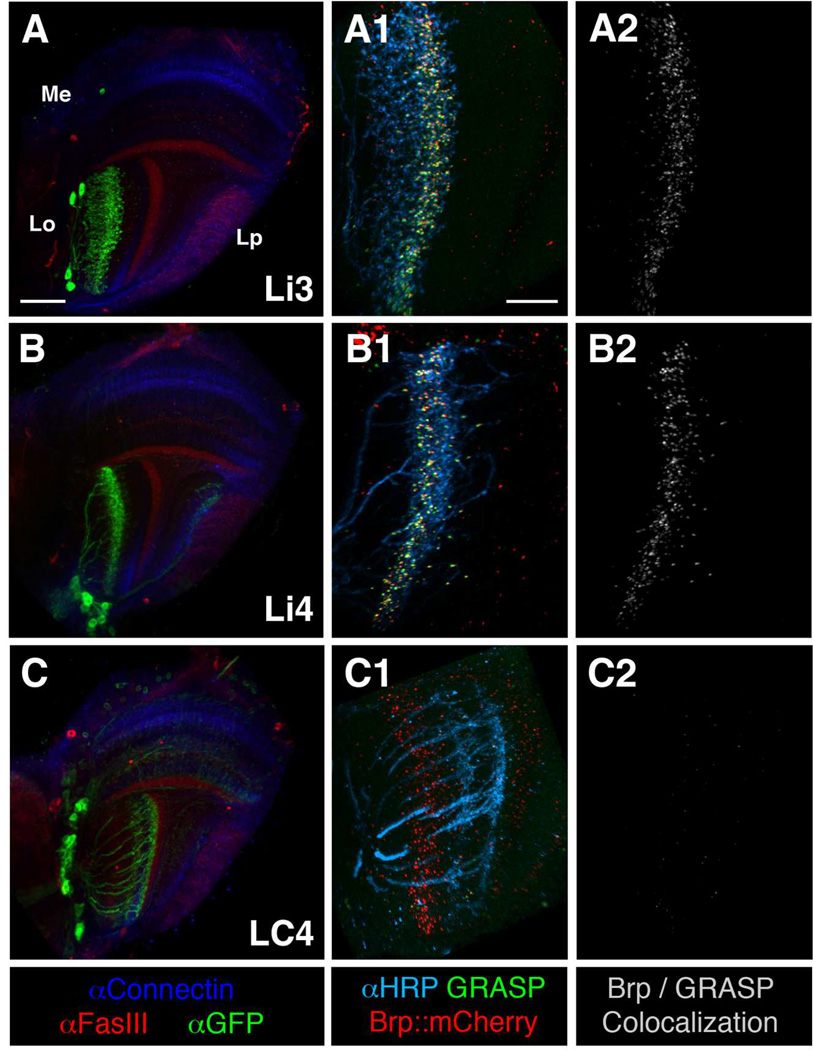
Figure 3.
Mapping chromatic lobula neurons by the GRASP method with presynaptic labeling. Lobula intrinsic neurons Li3 and Li4 receive densely packed synaptic contacts from the chromatic Tm neurons across lobula stratum 5. The lobula projection neuron LC4 forms very few or no synaptic contacts with the chromatic Tm neurons. Left panels (A–C): GAL4 lines used to express a membrane-tethered GFP marker (mCD8GFP, green) in three different types of lobula neurons (as indicated) to reveal their morphologies. FasIII (red) and Connectin (blue) antibodies labeling discrete medulla and lobula strata were used as layer-specific landmarks. Middle and right panels (A1-C2): ortC1a-LexA::VP16 drive expression of a split-GFP moiety (spGFP11-HA::CD4), and an active zone marker, BrpD3::mCherry, in four types of chromatic Tm neurons, Tm5a/b/c and Tm20. In the same animals, various lobula GAL4 lines express the other split-GFP (spGFP1-10::CD4) and the membrane marker HRP-CD2 in specific lobula neurons (as indicated). Functional GFP (GRASP) reconstituted at the membrane contacts of these two groups of neurons was examined for native fluorescence (green). Colocalizations (white) of the active zone marker (BrpD3::mCherry) and the GRASP signal indicated potential synaptic contacts (right panels, A2-C2). Lobula neurons are outlined by anti-HRP immunolabeling (cyan, middle panels A1-C1). Scale bar, 30 µm in A for A-C; 15 µm in A1 for A1-C2.

