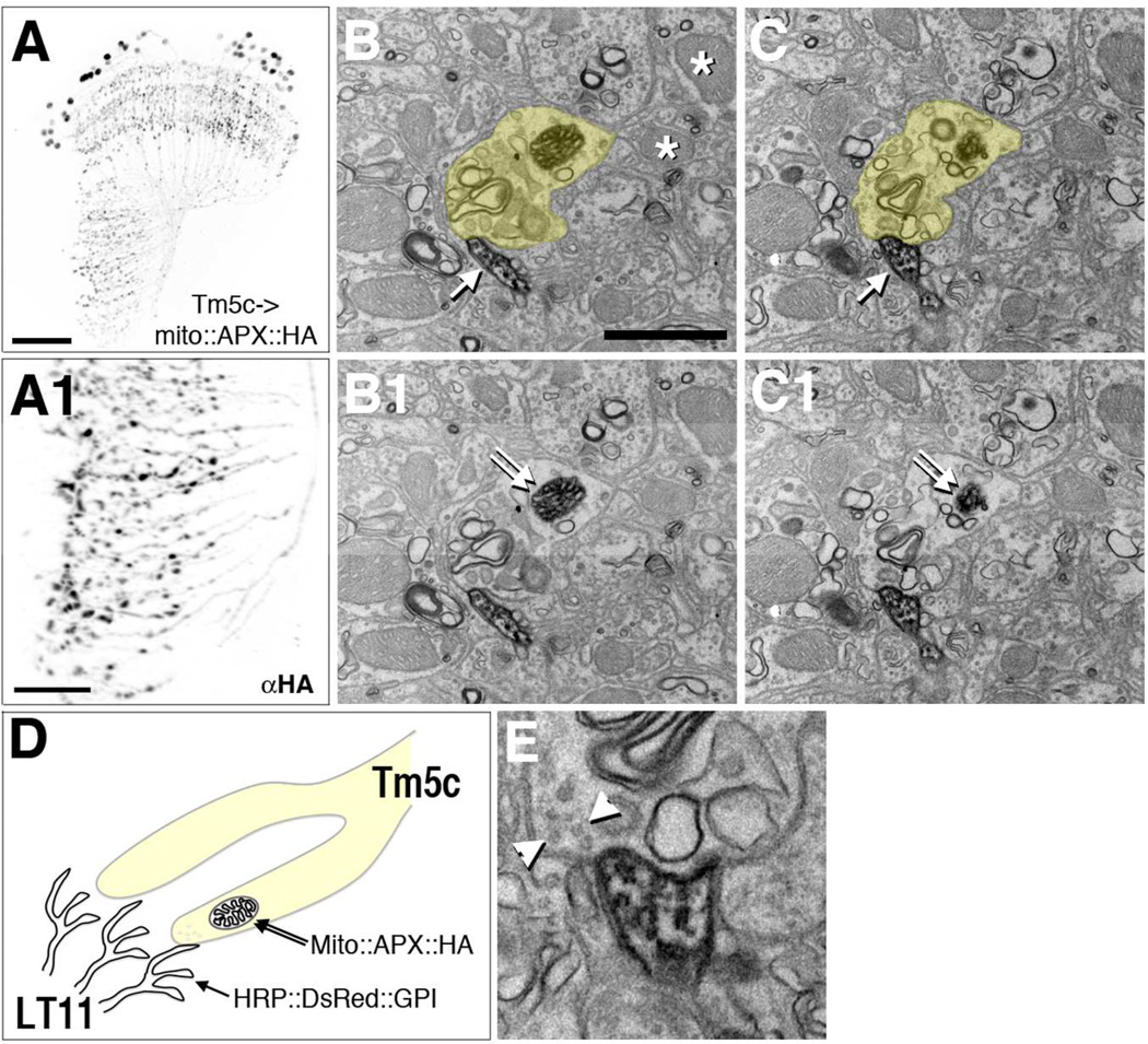
Figure 6.
“Two-tag” EM double labeling reveals Tm5c to LT11 contacts.
Tm5c neurons express HA-tagged APX targeted to the mitochondrion matrix (mito::APX::HA). Confocal microscopic analyses reveal Tm5c mitochondria labeled by anti-HA (αHA) in axon terminals in the lobula (A1) as well as dendrites and cell bodies (A). (A1) high magnification of the lobula of (A). Scale bar, 30 µm for A, 10 µm for A1.
Tm5c axon terminals identified by the presence of APX-labeled mitochondria and LT11 dendritic membrane labeled by membrane-tethered HRP (HRP::DsRed::GPI). (B-C1, E) EM sections of double-labeled lobula neuropil stained with DAB. A Tm5c mitochondrion identified by its dense DAB staining and mitochondrial morphology (double arrows, B1 and C1). Unlabeled mitochondria are indicated with asterisks (B). A Tm5c terminal (with a labeled mitochondria) is pseudocolored yellow in B and C, and raw images of the same area are presented in B1 and C1, respectively. LT11 dendrites identified by the DAB staining of the cytoplasmic membrane (single arrows in consecutive sections, B and C).
(D) A schematic illustration of “two-tag” double labeling method.
(E) High magnification view of the contact between Tm5c terminal and LT11 dendrites. Arrowheads indicate synaptic vesicles in Tm5c, compatible with the terminal being presynaptic to LT11 dendrites.

