Fig. 3.
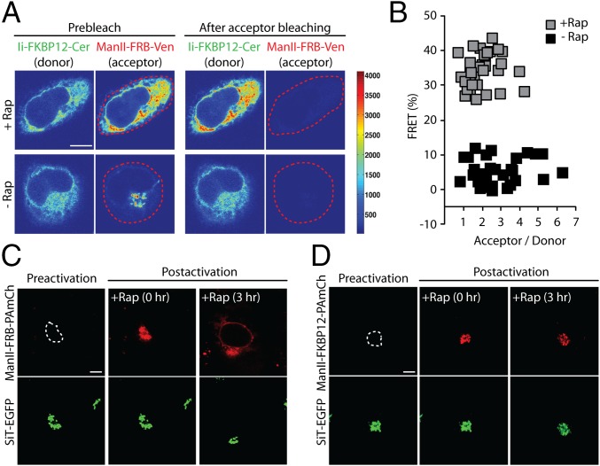
FRET and photoactivation-based assays confirm that redistribution of FRB-tagged Golgi enzymes to the ER is due to rapamycin-mediated complexation of recycling enzymes with an FKBP12-tagged ER trap. (A) HeLa cells expressing Ii-FKBP12-Cer and Man II-FRB-Venus were treated with cycloheximide ± rapamycin for 4 h. Confocal images of cells before and after photobleaching of the cellular Man II-FRB-Venus within the region indicated by the dashed line (red). (B) Percentage of FRET between Man II-FRB-Venus and Ii-FKBP12-Cer in HeLa cells imaged according to protocol described in A. (C) Golgi pool of Man II-FRB-PAmCh, transiently coexpressed in HeLa cells with Ii-FKBP12-Cer and SiT-EGFP, was selectively photoactivated within the region indicated by the dashed line. The Golgi region was identified by SiT-EGFP fluorescence. Images of a cell before photoactivation (preactivation), immediately after photoactivation (postactivation, 0 h), and after 3 h of incubation with cycloheximide ± rapamycin (postactivation, 3 h) (D) Golgi pool of Man II-FKBP12-PAmCh in HeLa cells, transiently coexpressed with Ii-FKBP12-Cer and SiT-EGFP, was selectively photoactivated within the region indicated by the dashed line. Images of a cell before photoactivation (preactivation), immediately after photoactivation (postactivation, 0 h), and after 3 h of incubation with cycloheximide ± rapamycin (postactivation, 3 h). (Scale bars: 10 μm.)

