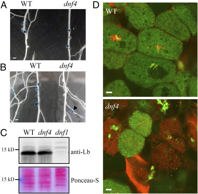
Fig. 2.
dnf4 nodules express fixation genes but fail to sustain differentiated bacteroids. (A and B) Activity of a nifH::GUS reporter in WT and dnf4 nodules at 10 dpi (A) and 14 dpi (B). The arrowhead in B denotes a young dnf4 nodule on a lateral root. (Scale bar: 1 mm.) (C) Accumulation of leghemoglobin in 10-dpi nodules. Ponceau S-stained membrane is shown as a loading control. (D) Live/dead staining of bacteroids in dnf4 compared with WT nodules (14 dpi). Live bacteroids have a green signal (GFP) and dying bacteroids have a red signal from propidium iodide (PI). PI can only enter bacteroids with compromised membranes. (Scale bar: 10 µm.)

