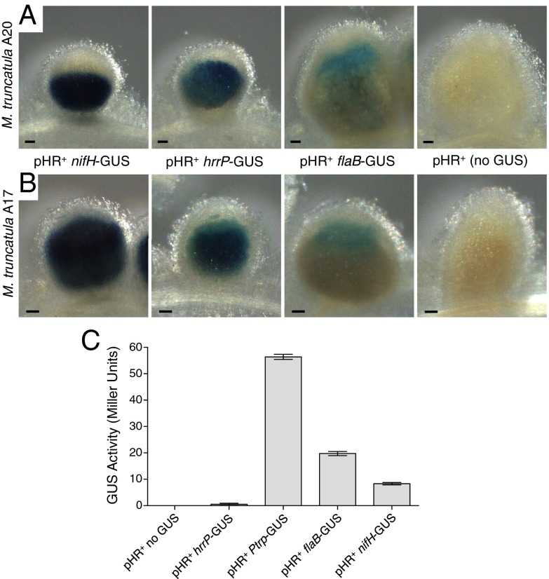
Fig. 3.
GUS promoter fusion studies showing nodule-specific expression of hrrP. (A and B) Representative micrographs of 10-d-old M. truncatula accession A20 (A) and A17 (B) nodules stained with X-GLUC to reveal the location and relative expression of the GUS reporter fusions integrated downstream of the nifHDK, hrrP, and flaB promoters (strains PP404, PP389, and PP397, respectively). All samples were stained under identical conditions. Wild-type B800 (no GUS) was used as a negative control for GUS straining in planta. (C) GUS expression levels in free-living cells grown to midlog phase. The constitutive Salmonella-derived trp promoter was included as a positive control (PP471). (Scale bars: 100 μm.) Error bars indicate SEM.

