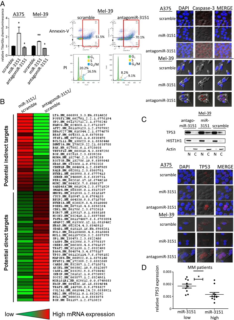
Fig. 1.
Effect of antagomiR-3151 on cell viability and discovery of miR-3151 downstream targets in MM. (A, Left) Viability of A375 and Mel-39 cells after forced miR-3151 expression or knockdown in chemiluminescent assays. Three experiments, bars, mean ± SD; *P < 0.05, **P < 0.005, two-tailed t test. In both A375 and Mel39 cells, overexpression of miR-3151 led to increased chemiluminescent activity and, thus, increased proliferation, whereas knockdown of miR-3151 led to decreased proliferation. (A, Middle) Example plots of annexin-V apoptosis assays and example histograms of propidium iodide cell cycle analysis. Green area is G1 phase, yellow is S phase, and blue is G2/M phase. Assays were performed in Mel-39 cells after miR-3151 knockdown. Infection with antagomiR-3151 led to an increase in the number of cells undergoing apoptosis and a decrease in cells undergoing cell division. (A, Right) Confocal microphotographs show A375 and Mel-39 cells transfected with scramble, miR-3151, or antagomiR-3151 stained for Caspase-3 (red). DAPI nuclear staining is shown in blue. In both cell lines, infection with antagomiR-3151 increased the expression of Caspase-3. (B) Heatmap of gene expression changes concordantly affected by forced miR-3151 expression or miR-3151 knockdown based on targeted RNA sequencing. Genes with concordant decreased expression upon miR-3151 overexpression and decreased expression after miR-3151 knockdown were considered potential direct targets of miR-3151. (C, Upper) Western blot with nuclear fractionation lysates to determine the relative nuclear (N) and cytoplasmic (C) abundance of TP53 in Mel-39 cells after manipulation of miR-3151 expression. Infection with antagomiR-3151 led to increased nuclear localization of TP53. (C, Lower) Confocal microphotographs show A375 and Mel-39 cells transfected with scramble, miR-3151, or antagomiR-3151 stained for TP53 (red). DAPI nuclear staining is shown in blue. When infected with antagomiR-3151, cells displayed increased nuclear localization of TP53. (D) TP53 expression in high miR-3151 and low miR-3151 expressing MM patients, *P < 0.05, two-tailed t test. Patients with high expression of miR-3151 had relatively lower TP53 expression compared with low miR-3151 expressers (as defined by median cut).

