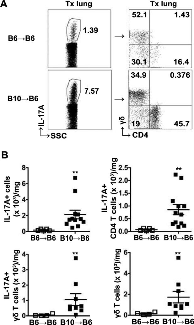
Figure 2. Increased IL-17A+ cells in allografts are CD4+ T cells and γδ T cells.
Transplanted lungs from B6→B6 and B10→B6 transplants were harvested at day 21 post-transplant and analyzed by flow cytometry. (A) Representative dot plots indicating frequency of IL-17A+ cells (left) of total lymphocytes and frequency of CD4+ T cells and γδ T cells (right) in IL-17A+ lymphocytes in transplanted left lung. (B) The absolute numbers of IL-17A+ lymphocytes, IL-17A+CD4+ T cells, IL-17A+γδ T cells and total γδ T cells in transplanted left lung. Absolute number of cells is normalized to the weight of the lung. For IL-17A+ cells and IL-17A+CD4+ T cells, n=5 for B6→B6, n=12 for B10→B6; for IL-17A+γδ T cells and total γδ T cells, n=4 for B6→B6, n=9 for B10→B6; data analyzed by Mann-Whitney U test, ** P<0.01.

