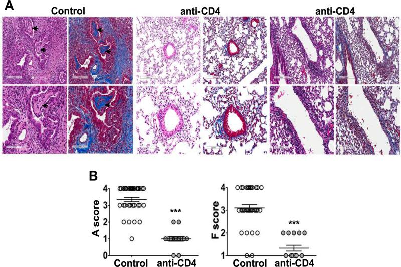
Figure 3. CD4+ T cells are required for rejection and the development of OB.
B6 recipients of B10 left lungs were treated with CD4-depleting mAb (GK1.5) two days prior to transplant and twice a week post-transplant, lungs were harvested and analyzed on Day 21. Untreated B10→B6 allografts were used as control. (A) H&E and Masson's trichrome staining of lung sections with no evidence of rejection in anti-CD4 treated allografts (middle panels) compared to cellular rejection and OB (arrow) found in the control allografts (left panels), an anti-CD4 treated allograft with mild cellular rejection and fibrosis has also been shown (right panels). magnification, 10X and 20X. (B) Acute rejection (A) scores and fibrosis (F) scores in transplanted left lung as described in methods, n=39 (control) and n=15 (anti-CD4), ***P<0.001 with Mann-Whitney U test. H&E, hematoxylin and eosin.

