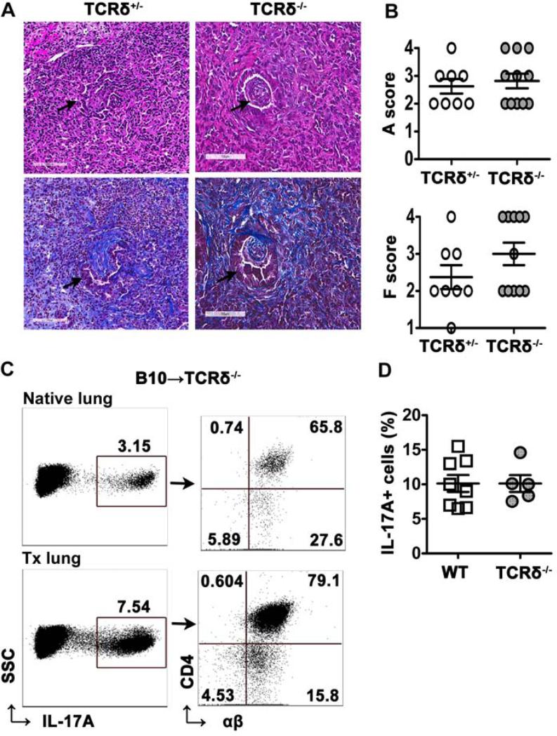
Figure 7. γδ T cell-deficiency does not prevent OB.
Left lungs from B10 mice were transplanted into TCRδ−/− or TCRδ+/− mice and analyzed at day 21 post-transplant. (A) H&E (top) and Masson's trichrome (bottom) stained lung allografts from TCRδ+/− and TCRδ−/− mice demonstrated OB-like lesions (arrows) in the airway, magnification, 20X. (B) Analysis of acute rejection (A) scores and fibrosis (F) scores in left lung allografts as described in methods, n=8 (TCRδ+/−), n=11 (TCRδ−/−). (C) Representative dot plots showing percentage of IL-17A+ lymphocytes on gated lymphocytes (left) and percentage of CD4+TCRβ+ T cells of IL-17A+ lymphocytes (right) in native lungs and transplanted left lungs from TCRδ−/− mice (n=5). (D) Frequency of IL-17A+ lymphocytes in transplanted left lungs from wild-type mice (n=7) as in Figure 4 and TCRδ−/− mice.

